
London Regional Microscopy Facility
Already have a Logon Account?
|
Welcome
Welcome to Robarts Research Institute, Western University FBS Portal. This site is designed to automate the use of our Core Facilities and to provide the best possible customer service. Quick Info
For more info, please contact the Priority Software Support Team. | Our Core Facilities To learn more about a particular facility or to request access, please click on a facility name below. Robarts Research Institute |
Main Contact Info
Claudia Seah or Suzanne Brett Welsh
Robarts Research Institute / Western University
1151 Richmond Street North
London, ON N6A 5B7
Remittance Contact Info
| Other Contacts | |||
|---|---|---|---|
| Paulina Kowalewska | VITAL Core Contact | (519) 931-5777 | vital-core@uwo.ca |
| Claudia Seah | Confocal Core Technical Manager | cseah@robarts.ca | |
This facility has not published any Products. Please check back.
The following Products and Services are available within our facility:
Analysis
 |
Leica Analysis WorkstationLeica Workstation |
Leica Aperio Analysis Workstation |
|
Nikon Image Analysis Workstation |
|
SP8 Analysis Only |
|
Zeiss Analysis Only |
Instruments
Evos Thermo-3260B |
|
 |
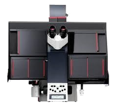
Leica TCS SP8-3281Leica TCS SP8 - Capabilities: |
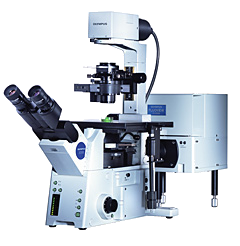 |
Olympus FV1000Olympus FV1000 - Capabilities: |
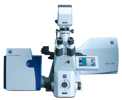 |
Zeiss LSM Meta 510-3260BZeiss LSM Meta 510 Capabilities: |
Olympus-Multi-Modal intravital |
|
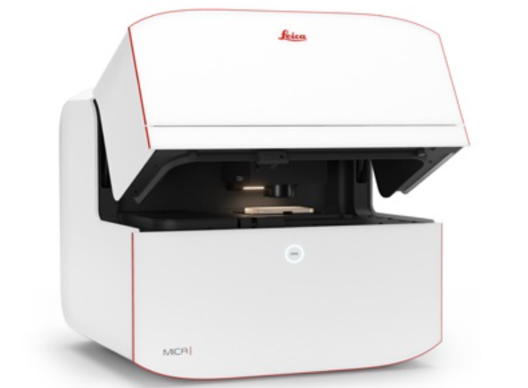 |
Leica SR GSD-3260DMICA is a highly automated widefield microscope. With the simple push of a button, you have everything you need - all in one place - to supercharge fluorescence imaging workflows and get meaningful scientific results faster. |
 |
Leica AM TIRF MC-3281Leica AM TIRF MC - Capabilities: |
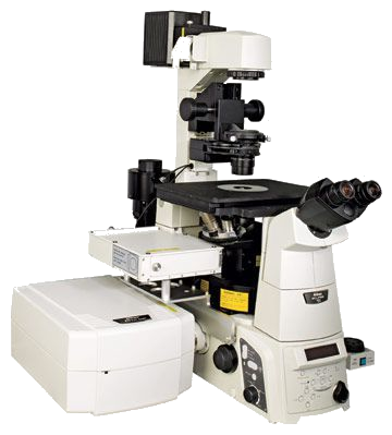 |
Nikon-Confocal-3277AThe Nikon A1R laser scanning confocal microscope fitted with Optisplit Bypass and Hamamatsu Flash 4 digital camera will have the same but much improved capabilities as the Olympus IX81 Inverted microscope. It also has a high-definition resonant scanner and a galvano scanner to capture fluorescence images of cells and structures in live animal preparations. |
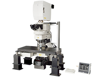 |
Nikon-Multiphoton-3277BThe multiphoton upright microscope with imaging capture computer can be used to image fluorescent images but in deeper tissues. Cells and tissues can be imaged at a greater depth than is currently possible with the inverted microscopes. |
Olympus-Multi-Modal intravital |
|
I-Stat Point Of Use Blood Analyzer |
|
Moor-Laser-Doppler |
Restricted Equipment & Analysis
Imaris Image Analysis SoftwareRestricted access. Imaris Software Package is an interactive visualization and analysis software for 3D and time-lapse microscopic images with advanced solutions for big datasets (Terabyte range) - smart Volume and Surface rendering based on our multi-resolution technologies, plus Surface calculations for images of unlimited size. |
|
 |
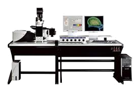
BrainScan-Stereology-System-Restricted UseRestricted access. |
Nanoflow-Restricted Use-3281Restricted access. Restricted equipment must be booked by users to accommodate COVID distancing |
This facility has not published any News. Please check back.
Quick Quotes have not been configured. Please check back soon (Code 001, Code 002)
, please enter that email address here.")